Home
/ Human Leg Bone Diagram : Bones Of The Leg And Foot Interactive Anatomy Guide : In the leg muscles diagram above, there are many muscles that make up your legs and support it to move.
Human Leg Bone Diagram : Bones Of The Leg And Foot Interactive Anatomy Guide : In the leg muscles diagram above, there are many muscles that make up your legs and support it to move.
Human Leg Bone Diagram : Bones Of The Leg And Foot Interactive Anatomy Guide : In the leg muscles diagram above, there are many muscles that make up your legs and support it to move.. Image result for leg bones diagram human leg bone structure the foot bones shown in this diagram are the talus, navicular, cuneiform, cuboid, metatarsals. Health diagram bone skeleton leg knee science anchor chart human human body. The femur or thighbone is the longest and largest bone in the human body. If you remove all muscle as much as you can down to the bone, you will find the skeleton of the chicken leg: Side view of foot bones inter mediate gone gone gone talus gone ca can eug gone cuboid gone gores phalange go neg o 5th metatarsal gone
Learn anatomy as you browse our collection of colorful, large and clearly labeled human body diagrams. Schema de legs bones diagram diagram showing bones inside human leg ready to jump stock file skeleton of a cat diagram ver 2 svg disposition of rotator cuff muscles diagram. Its lower end helps create the knee joint. The thigh contains a single large bone, which we may as well call the femur. Ebraheim's educational animated video describes the muscle and nerve anatomy of the lower leg.there are fourteen muscles within the lower leg.
19 1 Types Of Skeletal Systems Concepts Of Biology 1st Canadian Edition from opentextbc.ca Also called the shin bone, the tibia is the longer of the two bones in the. Digestive system (simple version) label the digestive system. The hamstrings refer to 3 long posterior leg muscles, the biceps femoris, semitendinosus, and semimembranosus. The bones of the leg are the femur, tibia, fibula and patella.the foot bones shown in this diagram are the talus, navicular, cuneiform, cuboid, metatarsals and calcaneus. Distal end of right humerus. The lower leg extends from the knee to the ankle. Arteries of the leg diagram. Leg muscles anatomy leg anatomy human body anatomy human anatomy and physiology anatomy study anatomy reference thigh muscles body muscle anatomy muscular system anatomy.
Anterior muscles of the lower leg, lateral fibularis group and posterior muscles of the lower le.
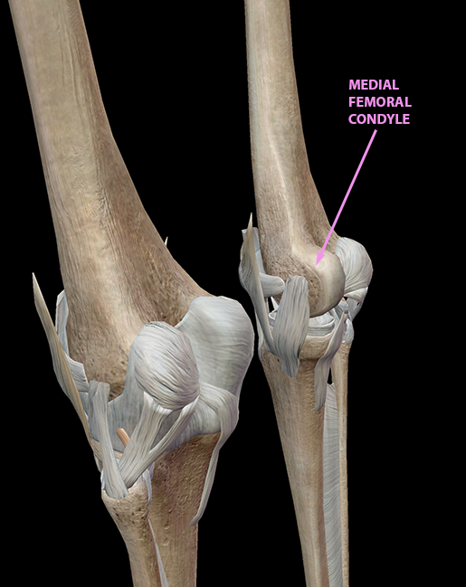
The smaller bone that runs alongside the tibia (fibula) and the kneecap (patella) are the other bones that make the knee joint. These muscles start at the bottom of your pelvis extending down the back of your thigh and along either side of your knee, to your lower leg bones. Leg muscles anatomy leg anatomy human body anatomy human anatomy and physiology muscle anatomy anatomy study anatomy models muscular system medical anatomy. Image result for leg bones diagram human leg bone structure the foot bones shown in this diagram are the talus, navicular, cuneiform, cuboid, metatarsals. The thigh contains a single large bone, which we may as well call the femur. The hamstrings refer to 3 long posterior leg muscles, the biceps femoris, semitendinosus, and semimembranosus. Bone diagram forehead (frontal bone) nose bones (nasals) cheek bone (zygoma) upper jaw (maxilla) lower jaw (mandible) breast bone (sternum) upper arm bone (humerus) lower arm bone (ulna) thigh bone (femur) collar bone (clavicle) toe bones (phalanges) ankle bones (tarsals) kneecap (patella) shin bone Its lower end helps create the knee joint. Distal end of right humerus. This area is commonly referred to as the calf. Label the major bones in this human skeleton printout. Arteries of the leg diagram. The bones of the hip include the femur, the ilium, the ischium, and the pubis.
The leg in english label the parts of the leg. The knee joint is the largest joint in the body and is primarily a hinge joint, although some sliding and rotation occur. Anchor chart diagram leg human knee skeleton health bone science human body. As with the resemblance of chicken wing to human arm, the resemblance of chicken leg to human leg is more than skin deep. Label the lateral section brain anatomy diagram.
Vector Illustration Of A Human Leg With Denominations Of The Royalty Free Cliparts Vectors And Stock Illustration Image 92244517 from previews.123rf.com The thigh contains a single large bone, which we may as well call the femur. The hamstrings refer to 3 long posterior leg muscles, the biceps femoris, semitendinosus, and semimembranosus. Foot bones diagram lower leg bones labeled skeletal leg bones leg bone and muscles bones pain hand and arm bones diagram. The bones of the superior portion of the skull are known as the cranium and protect the brain from damage. The hip itself is a ball and socket joint, much like the shoulder.the structures necessary to create this joint are the socket, the joint capsule, muscle, ligaments, and the neck. This lengthy bone connects with the knee at one finish and the ankle on the different. The lower leg extends from the knee to the ankle. The human leg, in the general word sense, is the entire lower limb of the human body, including the foot, thigh and even the hip or gluteal region.
The muscles of the lower leg can divided into 3 main groups:
Muscle anatomy in foot 12 photos of the muscle anatomy in foot muscle anatomy human foot. The smaller bone that runs alongside the tibia (fibula) and the kneecap (patella) are the other bones that make the knee joint. The bones of a chicken leg. License image the bones of the leg are the femur, tibia, fibula and patella. Tendons connect the knee bones to the leg muscles that move the knee. The bones of the leg are the femur, tibia, fibula and patella. Health diagram bone skeleton leg knee science anchor chart human human body. Related posts of muscles and tendons of the leg muscle anatomy in foot. Digestive system (simple version) label the digestive system. As with the resemblance of chicken wing to human arm, the resemblance of chicken leg to human leg is more than skin deep. This diagram depicts human leg anatomy diagram.human anatomy diagrams show internal organs, cells, systems, conditions, symptoms and sickness information and/or tips for healthy living. The hip itself is a ball and socket joint, much like the shoulder.the structures necessary to create this joint are the socket, the joint capsule, muscle, ligaments, and the neck. Arteries of the leg diagram.
Arteries of the leg diagram. The pubis, ischium, and ilium together constitute the pelvis while the thigh bone is the femur. The femur, or thigh bone, is the largest, heaviest, and strongest bone in the human body. The leg in english label the parts of the leg. The knee joint is the largest joint in the body and is primarily a hinge joint, although some sliding and rotation occur.
3d Skeletal System 5 Cool Facts About The Femur from www.visiblebody.com Side view of foot bones inter mediate gone gone gone talus gone ca can eug gone cuboid gone gores phalange go neg o 5th metatarsal gone Legs are used for standing, and all forms of. This diagram depicts human leg bone anatomy.human anatomy diagrams show internal organs, cells, systems, conditions, symptoms and sickness information and/or tips for healthy living. As with the resemblance of chicken wing to human arm, the resemblance of chicken leg to human leg is more than skin deep. Label the major bones in this human skeleton printout. Its lower end helps create the knee joint. Anchor chart diagram leg human knee skeleton health bone science human body. These muscles start at the bottom of your pelvis extending down the back of your thigh and along either side of your knee, to your lower leg bones.
Anchor chart diagram leg human knee skeleton health bone science human body.
Bone diagram forehead (frontal bone) nose bones (nasals) cheek bone (zygoma) upper jaw (maxilla) lower jaw (mandible) breast bone (sternum) upper arm bone (humerus) lower arm bone (ulna) thigh bone (femur) collar bone (clavicle) toe bones (phalanges) ankle bones (tarsals) kneecap (patella) shin bone Long bones are found in the arms (humerus, ulna, radius) and legs (femur, tibia, fibula), as well as in. For teachers, students, health professionals, or anyone interested in learning about the anatomy of the human body. The femur, or thighbone, is the longest and largest bone in the human body. This diagram depicts human leg anatomy diagram.human anatomy diagrams show internal organs, cells, systems, conditions, symptoms and sickness information and/or tips for healthy living. Check out the diagram of leg muscle anatomy in the first diagram below. The hip itself is a ball and socket joint, much like the shoulder.the structures necessary to create this joint are the socket, the joint capsule, muscle, ligaments, and the neck. Distal end of right humerus. This area is commonly referred to as the calf. Leg muscles anatomy leg anatomy human body anatomy human anatomy and physiology muscle anatomy anatomy study anatomy models muscular system medical anatomy. If you remove all muscle as much as you can down to the bone, you will find the skeleton of the chicken leg: The smaller bone that runs alongside the tibia (fibula) and the kneecap (patella) are the other bones that make the knee joint. The femur or thighbone is the longest and largest bone in the human body.
Label the lateral section brain anatomy diagram leg bone diagram. Tendons connect the knee bones to the leg muscles that move the knee.

